


[Event Report] The 110th HGPI Seminar – Deepening Industry-Academia Collaboration on iNPH Measures and Steps for Real-World Implementation (December 16, 2022)
date : 1/23/2023
Tags: Dementia, HGPI Seminar

![[Event Report] The 110th HGPI Seminar – Deepening Industry-Academia Collaboration on iNPH Measures and Steps for Real-World Implementation (December 16, 2022)](https://hgpi.org/en/wp-content/uploads/sites/2/hs110-top-1.jpg)
For our 110th HGPI Seminar, Health and Global Policy Institute (HGPI) hosted Dr. Shigeki Yamada of Nagoya City University Graduate School for a lecture on the current situation surrounding idiopathic Normal Pressure Hydrocephalus (iNPH), clinical studies involving industry-academia collaboration on AI-based automatic image recognition technology and gait disturbance detection, and issues and future prospects for implementing the use of such technology in society.
Key points of the lecture
- Idiopathic Normal Pressure Hydrocephalus (iNPH) is a form of dementia that is said to be undetected and undiagnosed in a significant number of people. Since iNPH can improve with treatment, it is referred to as a “treatable dementia.”
- In addition to cognitive impairment, the main symptoms of iNPH include gait disturbance and urinary incontinence. It is a progressive disease, and reports show that the risk of mortality triples if it is left untreated.
- A condition called Disproportionately Enlarged Subarachnoid-Space Hydrocephalus (DESH) is a key neuroimaging feature of iNPH and must be detected so the condition does not go overlooked.
- Dr. Yamada is involved in a joint industry-academia initiative. That is researching 3D and 4D imaging analysis and using AI for the automatic detection of DESH.
- A similar joint initiative led to the launch of a free iPhone application that can be used to conduct quantitative gait assessments using smartphones.
- A new AI-based 3D gait analysis smartphone application that enables people to spot the symptoms of iNPH on the individual level before seeing a specialist has been developed and will lead to more accurate diagnoses and treatments.
iNPH may be the second most prevalent form of dementia, after Alzheimer’s disease
It is said that one out of every five people age 65 and over will develop dementia. It has been projected that in 2025, the number of people living with dementia in Japan will reach approximately 7.3 million. Alzheimer’s disease (which affects 50% of the population with dementia and is the most common form of dementia) presents memory impairment, day night reversal (insomnia), and behavioral abnormalities (such as delirium and violence). Dementia with Lewy bodies (which affects 20% of the population with dementia and is the second most common form of dementia) presents altered cognitive function, visual hallucinations, abnormal sleep behavior, Parkinson’s disease signs, and constipation.
In addition to frontotemporal dementia (5%) and vascular dementia (15%), another rare form of dementia is idiopathic Normal Pressure Hydrocephalus (iNPH), and this is the condition we want to address. People living with iNPH often present symptoms like lack of awareness toward surroundings, lack of motivation, easily forgetting tasks they have completed, and being easily distracted. It is estimated that iNPH accounts for 3% of all dementia cases. However, some believe the number of potential patients whose iNPH is undiagnosed may be ten times higher. If iNPH actually accounts for 30% of all cases of dementia, it may be the second most common cause of dementia after Alzheimer’s disease.
How iNPH symptoms progress
Among the symptoms characteristic of iNPH, the disease is more often detected by gait disturbances and falls than by declines in cognitive function. The gait that is characteristic of iNPH begins to manifest as a wobble then includes shuffling, a small stride, a wide base. It becomes difficult for the affected person to turn and easier for them to fall. They may be unable to stop advancing when walking on a downward slope or experience frozen gait, which prevents them from taking a step. These also increase the risk of falls. Once someone experiences a fall, walking becomes frightening. This leads to fewer opportunities to walk alone, resulting in weaker leg muscles and further declines in activity, which in turn compounds the decline in cognitive function. A recent study from Sweden reported that the risk of mortality is tripled if iNPH goes untreated, and the world is beginning to view it as “a disease that ends in death.”
Providing information online with the company
For some time, iNPH has been known as a “treatable dementia,” but since it is easier to detect iNPH by gait disturbance and falls than by declines in cognitive function, and because the effects of treating gait disturbance are clearer, activities to raise awareness for iNPH are centered around “preventable falls.” A website titled “Hydrocephalus in Elderly People iNPH.jp” includes stories from people with iNPH and their families, essays from physicians, and similar content. I am also collaborating on that initiative.
What is DESH?
When the Guidelines for Management of Idiopathic Normal Pressure Hydrocephalus (Second Edition) were published in 2011, one of its new additions was to point out the importance of conducting imaging studies to detect Disproportionately Enlarged Subarachnoid-Space Hydrocephalus (DESH). The third edition was published in 2020 and included additional content on the impact of the key comorbidities of iNPH and methods of reducing shunt procedure complications (including shunt and valve management, postoperative outcomes, long-term management, and rehabilitation).
In addition to ventricular enlargement, the neuroimaging features of DESH include unbalanced cerebrospinal fluid (CSF) distribution in the subarachnoid space, an enlargement of the Sylvian fissure, and narrowing of the sulci over the high-convexity/midline surface. After detecting DESH in neuroimaging studies, a CSF tap test is performed in patients who clearly exhibit the typical triad of iNPH symptoms: gait disturbance, cognitive dysfunction, and urinary incontinence (including frequent urination and lack of awareness of urinary urge). When it is confirmed that symptoms have improved, a spinal shunt is often indicated.
Normally, CSF is driven by cerebral circulation to maintain consistent intracranial pressure. CSF movement is believed to be related to ventricular enlargement and DESH as they manifest in iNPH, and imaging studies on this are now advancing. A new method called 4D Flow MRI can be used to measure CSF flow as it is driven by the beating of the heart. In healthy people, more CSF enters the ventricles as CSF increases as the brain atrophies with age and as the foramen of Magendie (the boundary between the ventricles and the subarachnoid space) widens. This makes it easier for CSF pulsations to propagate into the ventricles during cerebral circulation. I believe these increased CSF pulsations may damage the ventricle walls, causing them to enlarge. People with iNPH have even stronger CSF pulsations within the ventricles than healthy people at the same age, which results in abnormal ventricular enlargement.
Evaluating DESH (by degree of severity) and joint industry-academia research
Several methods have been proposed for evaluating DESH in terms of the degree of sulcus narrowing in the high convexity and Sylvian fissure enlargement. These include a method of grading each one on a four-point scale (from 0 to 3) (Narita et al. 2016); the iNPH Radscale, which is widely used in Europe and grades narrowing of high parietal sulci using three levels (0 to 2) and Sylvian fissure enlargement with two levels (present or not) (Kockum et al. 2018). Both methods are basically subjective evaluations.
Subjective evaluation methods are superior in terms of simplicity, but DESH is difficult to detect for inexperienced physicians who do not routinely view images. There is also the problem that even experts are divided on evaluations. I discovered that in iNPH, ventricles tend to expand more in the Z-axis (vertically) than in the X-axis (horizontally). As such, I think the Evans index, a well-known index of ventricular expansion, is not suitable for evaluating ventricular enlargement in iNPH because it measures width along the X-axis. I have proposed a new index which measures along the Z-axis called Z-Evans index as an indicator of ventricular enlargement in iNPH (Yamada et al. 2015).
Professor Kazunari Ishii of Kindai University has also proposed an index in which the callosal angle of the ceiling of the lateral ventricles becomes steep (less than 90 degrees) as a result of ventricular enlargement along the Z-axis (Ishii et al. 2008). This index has been adopted worldwide. Furthermore, I have discovered that brain compression above the ventricles due to ventricular enlargement along the Z-axis is a key neuroimaging feature of iNPH, and have proposed a Brain/Ventricle Ratio (BVR) at AC<1.0 and a Convexity-SAS/Ventricle Ratio (CVR) of <1.0 as discrimination indices for DESH.
It has recently become possible to take 3D images of the brain with MRIs. Using a 3D medical imaging analysis system from the company, who I am conducting joint research with, it is possible to automatically split the brain into 21 regions (such as the frontal, temporal, parietal, and occipital lobes) and CSF in five regions (including each ventricle and subarachnoid space) and instantly calculate the volume of each. Utilizing an application for analyzing each region of the brain has made it easier to calculate 3D indices like CVR.
Our joint industry-academia research initiative on imaging analysis uses a cloud-based AI diagnostic imaging development support service, that initiative has succeeded in developing an AI-based system for automatically detecting DESH, a key neuroimaging feature iNPH. It also instantly extracts regions and calculates volume in the ventricles, subarachnoid spaces in the high-convexity and midline, the Silvian fissure, and the basilar cistern. We are currently working to see the use of this system implemented in society.
Cerebrospinal Fluid (CSF) tap tests
In a CSF tap test, which is used to determine the need for a CSF shunt procedure, a lumbar puncture is performed with a spinal needle of a somewhat large diameter (19G) to slowly drain approximately 30 ml of CSF by spontaneous drainage. We then compare symptoms before and after the procedure to check if they improved.
If the patient’s symptoms improve, they are indicated for surgery. There are three kinds of shunts used in CSF shunt procedures: ventriculoperitoneal (VP) shunts, ventriculoatrial (VA) shunts, and lumbar-peritoneal (LP) shunts. To increase surgery success rates, I have recommended physicians use the VINCENT craniotomy simulator application (from Fujifilm Co, Ltd.) to simulate the unique shape of the patient’s head and ventricles before the procedure to determine the optimal ventricular puncture route.
Estimating post-shunt improvement in symptoms at the stage of CSF tap testing
Different people produce different results when evaluating improvements in symptoms like gait disturbance, cognitive dysfunction, and urinary incontinence. There are also differences among patients and their families regarding the minimum potential improvements they require before they are willing to undergo surgery. If there are significant gaps in expectations among physicians and patients, it can result in trouble after the procedure. In this manner, subjective evaluations and senses of value vary from person to person, so I think it is important to make objective evaluations by quantifying the severity of gait disturbance and cognitive dysfunction as well as prospects for improvement.
Since its first edition was published, the Guidelines for Management of Idiopathic Normal Pressure Hydrocephalus have recommended the three meter Timed Up & Go (TUG) test. The TUG test measures how many seconds a patient takes to stand up from a chair on cue, walk in a straight line for three meters one way, turn around, walk back in a straight line, turn around, and sit down again. Two nationwide multicenter studies called SINPHONI (which used VP shunt procedures) and SINPHONI-2 (which used LP shunt procedures) were conducted to generate estimates of how many seconds patients could be expected to improve on the TUG test after shunt procedures according to their TUG time after a CSF tap test. They found that when CSF tap tests improved TUG test results by 5 seconds or more, at one year, both VP and LP shunt procedures resulted in an improvement of 10 seconds or more for approximately 40% of people and at least 5 seconds or more for approximately 65% of people.
Most patients whose TUG times worsened or improved by less than five seconds after CSF tap tests were highly likely to see their TUG time worsen or improve by less than five seconds one year after a VP or LP shunt procedure. However, patients with mild gait disturbance whose original TUG times (before CSF tap test) were less than 20 seconds had little room for TUG time improvement after CSF tap tests and shunt procedures. This led us to feel that other objective assessment methods would be necessary.
Utilizing smartphone applications in gait analysis research
Against this backdrop, in 2017, we began a new industry-academia collaboration to develop a smartphone application for conducting gait analysis. Research is ongoing and now includes participation from the Tesseikai Spine and Spinal Cord Center, Yamagata University, the National Institute of Advanced Industrial Science and Technology (AIST), and Nagoya City University.
First, we conducted a gait analysis study to measure a smartphone application. This was installed on a smartphone that participants would hold in a pouch worn near the stomach. That study found CSF tap tests did not improve TUG times in people with iNPH and mild gait disturbances but did improve trunk acceleration in all three dimensions (namely, forward, vertical, and lateral acceleration). We also found that the 95% confidence ellipsoid volume of this 3D acceleration is useful as an indicator.
We assigned 50 points to TUG times of 13.5 seconds. For participants with relatively severe gait disturbance and TUG times greater than 15.5 seconds, we reflected TUG times; and for those with relatively mild gait disturbance and TUG times less than 13.5 seconds, we reflected the 95% confidence ellipsoid volume of 3D trunk acceleration. Results were then distributed from 0 to 100 to define Instrumented Timed Up and Go (iTUG) scores. Together with Mr. Yukihiko Aoyagi of Digital Standard Co, Ltd., we developed a smartphone application called “Hacaro Series iTUG” to instantly measure iTUG times (Yamada et al. 2019).
The objective of efforts to implement the smartphone application in society is not only to get as many people as possible to use it but to also encourage the adoption of iTUG times as an international standard for gait analysis. To this end, the application has been made available for free in both Japanese and English. In addition to hospitals and long-term care facilities throughout Japan, Hacaro Series iTUG is now being used in hospitals in South Korea and Europe.
In addition to evaluating the severity of gait disturbance, we wondered if we could also use the smartphone application to quantify the pathological gait patterns iNPH, such as shuffling, small stride, or wide-based gait. We had participants walk 15 paces in a straight line and analyzed changes in their forward, vertical, and lateral trunk acceleration. We found the pathological gait of iNPH is characterized by small variations in forward and vertical acceleration and large variations in lateral acceleration. However, we concluded that we could not discern gait patterns like shuffling, small stride, or wide-based gait by examining changes in trunk acceleration.
The Three-Dimensional Pose Tracker for Gait Test (TDPT-GT)
Motion capture is a well-known method for objectively evaluating gait patterns including shuffling, small stride, or wide-based gaits, but it requires very expensive equipment, large spaces, and for markers to be attached across participants’ entire bodies. These hurdles make it difficult to adopt motion capture technology across the fields of healthcare and long-term care. This led us to wonder if we could design a motion capture system using only a single smartphone in which participants did not have to wear any markers. This led our joint industry-academia research initiative to develop the Three-Dimensional Pose Tracker for Gait Test (TDPT-GT), a new smartphone application that provides 3D motion capture (Aoyagi et al. Sensors. 2022).
To develop TDPT-GT, we did not train an AI using actual human movement. Instead, we started by developing a 3D character that is stored in the computer and moves according to instructions. It is then filmed by a virtual camera to generate a large amount of data (over 1 million movement patterns) to train the AI. The character was given bone data (a skeleton) so the AI could instantly track the coordinates of each joint, which made it ideal for machine learning. Running this AI with the high-performance neural engine included in recent iPhone models makes it possible to use the iPhone camera to instantly estimate the 3D coordinates of 24 points on the body, from head to toe. For example, it can even convert the movements of a baseball player on a television screen into 3D motion.
A study to verify the reliability of the relative 3D coordinates obtained using the TDPT-GT smartphone application is currently being verified by Professor Yoshiyuki Kobayashi, who is a world leader in motion capture gait analysis and a member of the EXercise motivation and Physical function Augmentation Research Team (ExPAR) at the AIST Human Augmentation Research Center. Although changes in joint angles can be calculated using the relative 3D coordinates obtained with the TDPT-GT smartphone application, instead of using 3D angles, we thought it would be easier to understand various aspects of gait, such as leg angle, using 2D angles obtained by projecting 2D relative coordinates onto the body axis planes (namely, the sagittal, coronal, and axial planes). We then plotted movement trajectories along each projection plane to calculate the 75% confidence ellipse and then used the center point, length of the major axis, and maximum and minimum values of the ellipse to search for pathological gait indicators.
When measuring in two dimensions, we discovered that it was likely for the participant to have shuffling gait if the average angle range of the hip joint was less than 30% in the sagittal projection plane. For the knee joint, if the average angle range of the hip joint was less than 45% in the sagittal projection plane, they were likely to have shuffling gait and short-stepped gait. There was a high likelihood of shuffling gait when vertical amplitude of the left and right heels was less than 10% of the length of the lower legs. Looking at the lateral deviation of stance (how open their stance was), we also found that the likelihood of wide-based gait was high if lateral deviation in the toes relative to the heels or in the heels relative to the hip joints in the axial plane projection was 10% or more of the length of the lower leg. We would like to develop a new smartphone application that can instantly measure and display these indices and for it to achieve real-world implementation so shuffle gait, wide-based gait, short-stepped gait, and fall risk can be detected on the individual level without the need to visit a specialist in a hospital.
[Reference] Publications related to gait analysis using TDPT-GT
Quantitative Gait Feature Assessment on Two-Dimensional Body Axis Projection Planes Converted from Three-Dimensional Coordinates Estimated with a Deep Learning Smartphone App
Website
PDF Version
The potential of big data and health data
iPhones come with an application called “Apple Research” which allows users to participate in studies. For example, the Apple Heart and Movement Study being conducted in the U.S. has accumulated 175,000 registered participants, data from 33 million workouts, and 40,000 fall records. Moving forward, I think using big data like this will reveal new information on fall risk.
The six-minute walk test (6MWT) and 30 second chair stand test (30CST) are both considered vital in diagnosing iNPH, but with newer iPhone models, just holding one allows the device to gather number of steps, stride length, and double support time, which are all stored as health data. In addition, using an AI developed by Apple Research, these devices can also conduct a pseudo 6MWT and measure walking steadiness. We are now living in an era in which we can utilize health data to assess gait and fall risk before and after CSF tap tests and shunt procedures in patients with iNPH, and in which health data can be confirmed on the individual level.
The Stroop Test cognitive function assessment application
Two tests that are widely used to evaluate cognitive function are the Mini-Mental State Examination (MMSE) and the Revised Hasegawa’s dementia scale (HDS-R). These tests are also useful in iNPH treatment if conducted before and after CSF tap test and shunt procedures. However, iNPH has a specific form of cognitive decline in which people’s movements become slow or they forget things they have done. The Hellström Scale (which was developed in Europe in 2012) and the Stroop Color and Word Test (SCWT) are widely used to evaluate aspects of cognitive function, but the Stroop Test is not widely used in Japan and many hospitals do not have test equipment. So, I asked Digital Standard Co, Ltd. to release a free, simplified version of the Stroop Test as an application. This led to the release of the Hacaro Series Stroop Test, which is now being used in many hospitals to conduct evaluations before and after CSF tap tests and shunt procedures.
As we can see, we are now living in a time when ICT is being utilized in healthcare and long-term care services so people do not have to rely solely on the expertise of physicians. Instead of patients only being provided with diagnoses after visiting hospitals, I think it is important that we incorporate AI and smart devices into medical care so everyone, starting with patients and their families, can recognize symptoms and illnesses on their own and be able to seek more accurate diagnoses and treatments.
[Event overview]
- Speaker: Dr. Shigeki Yamada (Lecturer, Department of Neurosurgery, Graduate School of Medical Sciences, Nagoya City University)
- Date & time: Friday, December, 16, 2022; 18:30-20:00 JST
- Format: Online (Zoom Webinar)
- Language: Japanese
- Participation Fee: Free
- Capacity: 500
Speaker profile:
Dr. Shigeki Yamada (Lecturer, Department of Neurosurgery, Graduate School of Medical Sciences, Nagoya City University)
Dr. Shigeki Yamada was born in the City of Nagoya. After graduating from the Gifu University Graduate School of Medicine in 1997, he joined the medical office of the Kyoto University Department of Neurosurgery. In 2001, he began the doctoral program at Kyoto University Graduate School of Medicine and Faculty of Medicine, studying genetic analysis, medical statistics, and epidemiology. He began a collaborative research initiative with the University of Tokyo Institute of Industrial Science in 2002. In 2004, he attended the Centre National de Génotypage (the National Center of Human Genomics Research) in France. He earned his Doctor of Medicine degree in 2004, was named a board-certified neurosurgeon of the Japan Neurosurgical Society in 2005, and was certified as a medical specialist in stroke by the Japan Stroke Society in 2007. From 2017 to the present, Professor Yamada has been involved in joint industry-academia initiatives spanning the fields of medicine and engineering in which he is researching 3D image analysis with Fujifilm Co., Ltd. and 3D gait analysis with Digital Standard Co., Ltd. From 2013 to 2019, Professor Yamada studied under Dr. Masatsune Ishikawa at the Rakuwakai Otowa Hospital and developed a specialty in idiopathic Normal Pressure Hydrocephalus (iNPH). He served at the Department of Neurosurgery at the Shiga University of Medical Science from 2019 to 2022. In October 2022, he assumed a role at the Nagoya City University Department of Neurosurgery where he will be involved in research to raise awareness of iNPH, develop new diagnostic and therapeutic methods for iNPH, and conduct research to elucidate its pathophysiology.
日本語
Related Posts
-
2023-05-08
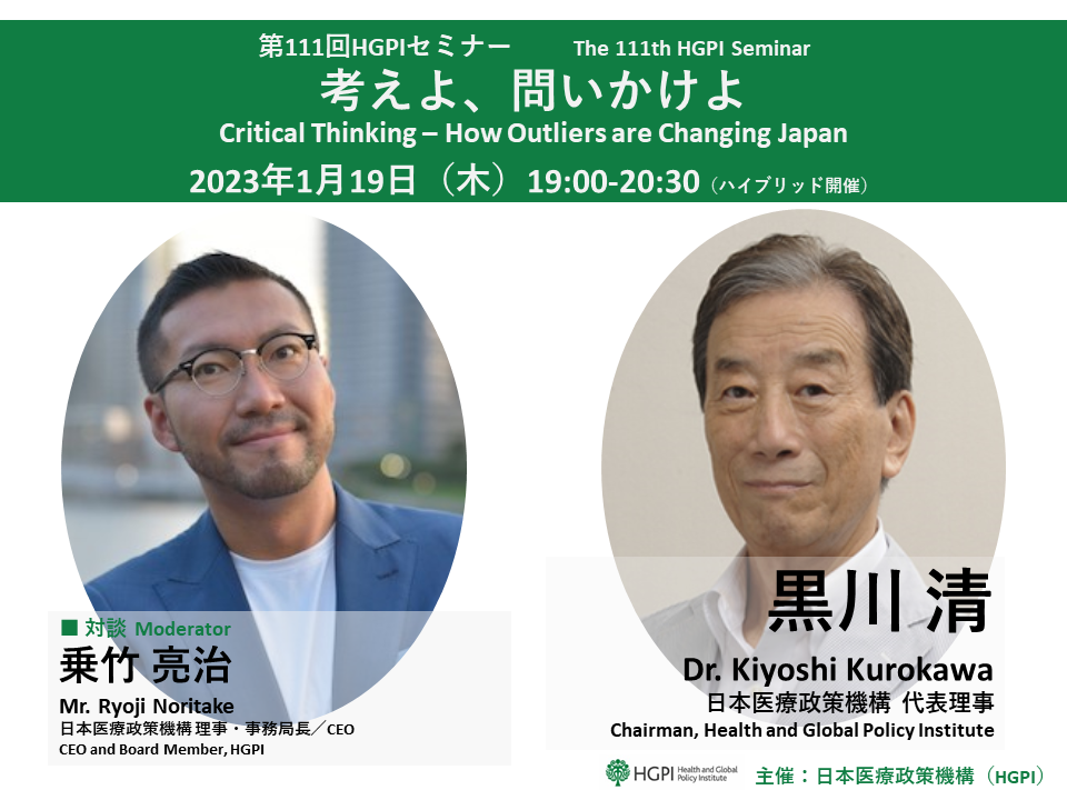
[Event Report] The 111th HGPI Seminar – Critical Thinking – How Outliers are Changing Japan (January 19, 2023)
-

2023-01-18
[Event Report] The 109th HGPI Seminar “The Clinical Treatment of Infectious Diseases in Small Animals Today and in the Future – Current Circumstances for AMR Control and Emerging Infectious Diseases in Companion Animals” (December 9, 2022)
-
2023-04-26
[Event Report] Non-partisan Diet Member Briefing – 30-minute Health Policy Update: “Introducing Idiopathic Normal Pressure Hydrocephalus (iNPH), a Form of Dementia That Can Improve With Treatment” (April 11, 2023)
-
2023-02-17
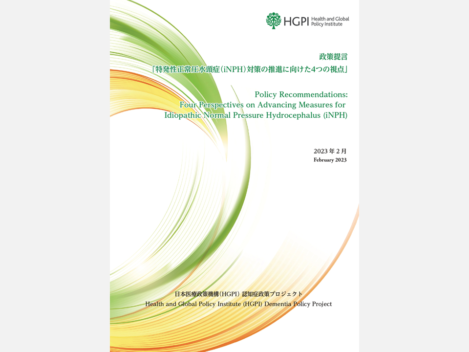
[Policy Recommendations] Four Perspectives on Advancing Measures for Idiopathic Normal Pressure Hydrocephalus (iNPH) (February 16, 2023)
-

2022-11-22
[Event Report] The Health and Global Policy Institute Dementia Policy Project Public Symposium: “Current Issues and Future Prospects for Idiopathic Normal Pressure Hydrocephalus (iNPH) Measures – Focusing on a Form of Dementia that Improves with Treatment” (August 24, 2022)
Top Research & Recommendations Posts
- [Report and Recommendations] Discussion Points in Healthcare DX Project Expert Panel Meeting (April 2, 2024)
- [Event Report] Planetary Health Expert Meeting Aiming for Sustainable Healthcare: Learning from the Impact of Environmental Pollution and Medical Waste During the Pandemic (February 16, 2024)
- [Announcement] A Significant Step Towards the Building a Green Healthcare System: Support for the Formal Expression of Interest by the Japanese Government Delegation to the ATACH at the Executive Board Meeting of the WHO (February 16, 2024)
- [Policy Recommendations] Kidney Disease Control Promotion Project 2023 “Establishing Kidney Disease Control Measures with Patient, Citizen, and Community Engagement and Collaboration” Policy Recommendations, a Collection of Good Practices of Chronic Kidney Disease (CKD) and Control Measures in Local Governments (February 14, 2024)
- [Research Report] Building a Mental Health Program for Children and Measuring its Effectiveness (June 16, 2022)
- [Research Report] 2019 Survey on Healthcare in Japan
- [Research Report] Survey of Japanese Physicians Regarding Climate Change and Health (December 3, 2023)
- [Policy Recommendations] Dementia Prevention Initiatives for Achieving a Dementia-Friendly and Inclusive Society (March 26, 2024)
- [Announcement] HGPI Joins Global Climate and Health Alliance (March 23, 2024)
- [Policy Recommendations] Three Necessary Perspectives for Formulating the Basic Plan for the Promotion of Policies on Dementia: Creating a Society That is Inclusive for All People at All Times (April 1, 2024)
Featured Posts
-
2024-04-12
[Announcement] HGPI Joins Global Climate and Health Alliance (March 23, 2024)
![[Announcement] HGPI Joins Global Climate and Health Alliance (March 23, 2024)](https://hgpi.org/en/wp-content/uploads/sites/2/ph-20240323-top.png)
-
2024-04-16
[Activity Report] HGPI Child Health Project Selected to Implement FY2024 Nippon Foundation Grant Program, “Establishing a Skill Development Program and Collaborative Network for Improving Mental Health for Students with Intellectual Disabilities” (April 15, 2024)
![[Activity Report] HGPI Child Health Project Selected to Implement FY2024 Nippon Foundation Grant Program, “Establishing a Skill Development Program and Collaborative Network for Improving Mental Health for Students with Intellectual Disabilities” (April 15, 2024)](https://hgpi.org/en/wp-content/uploads/sites/2/child-health-project.png)
-
2024-04-18
[Event Report] NCDs-related Cross-project Meeting: The 2nd Meeting on Non-Communicable Disease in Hokkaido and Tohoku (March 15, 2024)
![[Event Report] NCDs-related Cross-project Meeting: The 2nd Meeting on Non-Communicable Disease in Hokkaido and Tohoku (March 15, 2024)](https://hgpi.org/en/wp-content/uploads/sites/2/ncd-ckd-ob-cvd-20240315-top.jpg)
-
2024-04-23
[Registration Open] The 125th HGPI Seminar: Progress and Prospects for Domestic Measures on Health Problems Caused by Alcohol (May 24 , 2024)
![[Registration Open] The 125th HGPI Seminar: Progress and Prospects for Domestic Measures on Health Problems Caused by Alcohol (May 24 , 2024)](https://hgpi.org/en/wp-content/uploads/sites/2/HGPI_20240524_125thHGPIseminar_Eyecatch_rev.jpg)
